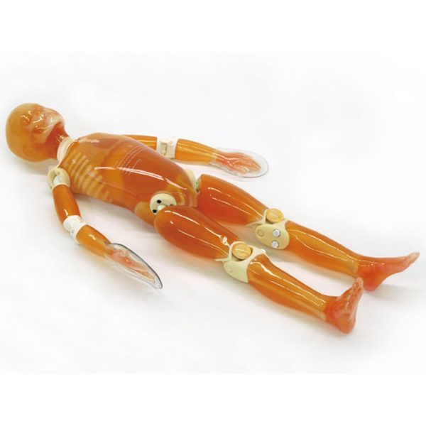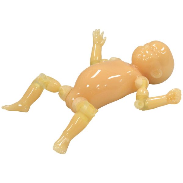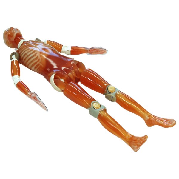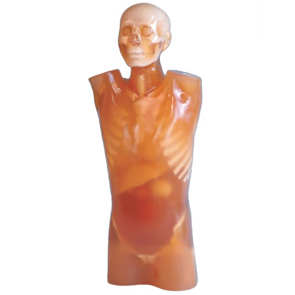
Features
- Design focused on positioning and light weight with clear images.
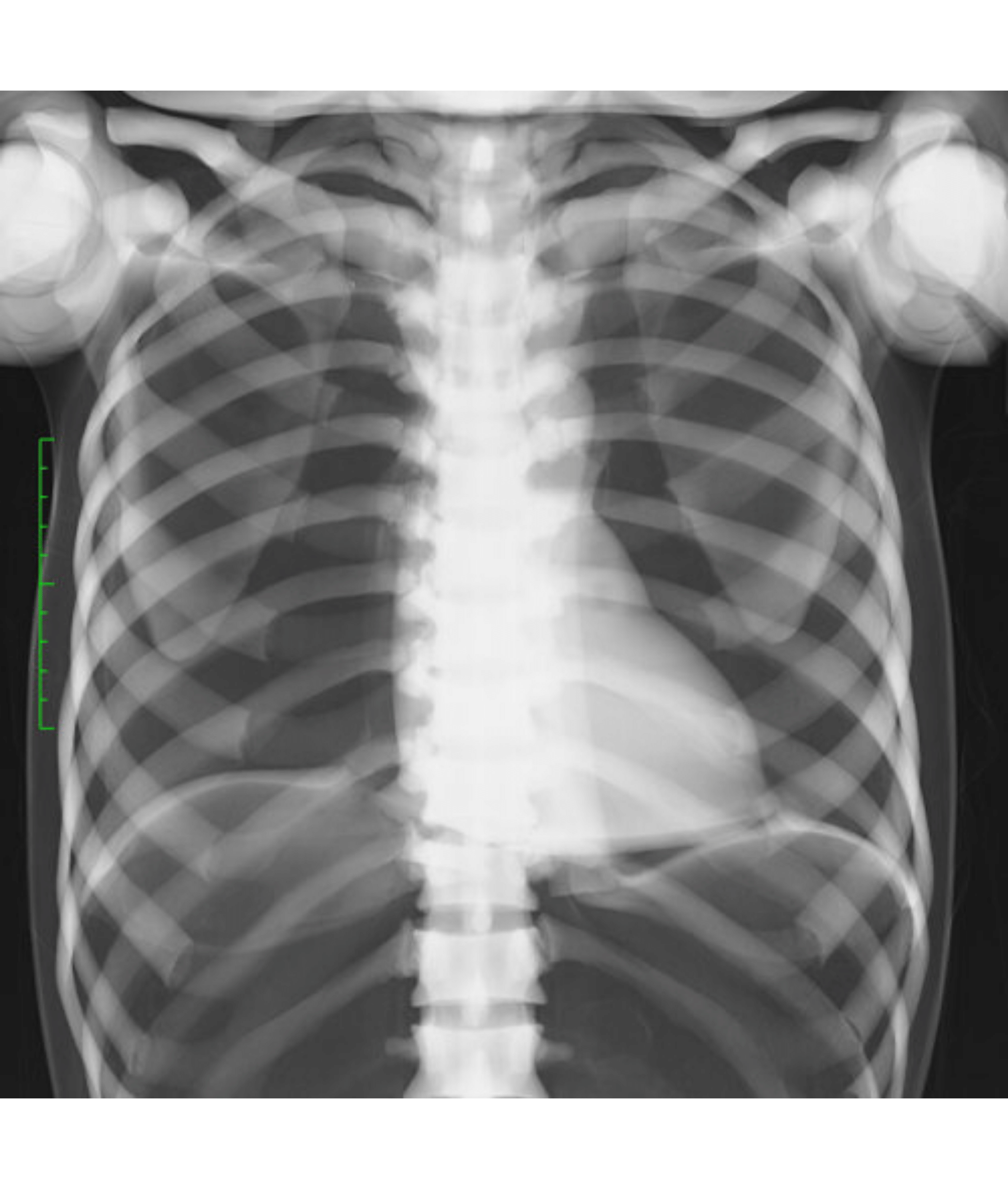
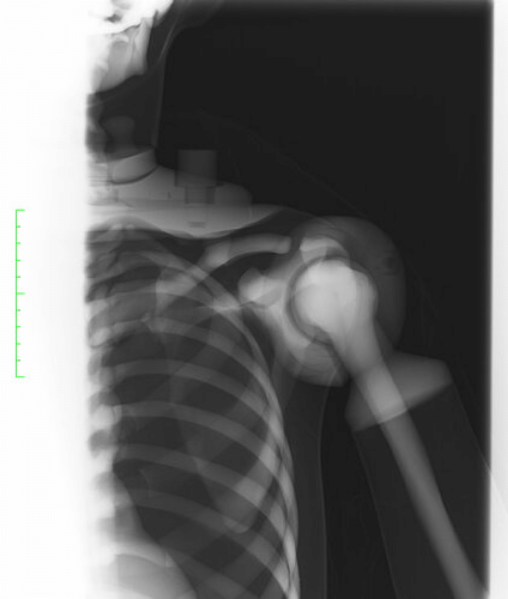
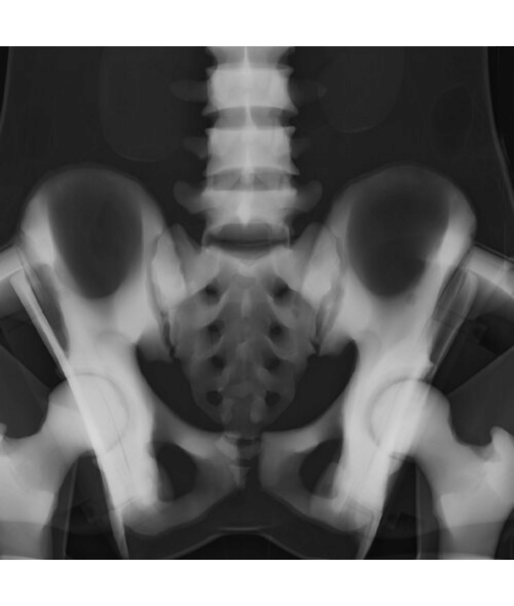
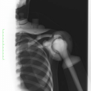
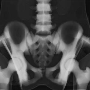
- Radiography images can be acquired with a lower irradiation than the real one and reduce the radiation exposure to trainees and stress on the device.
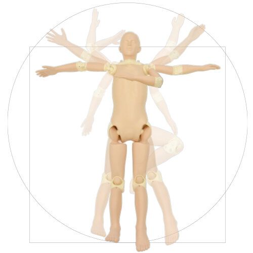
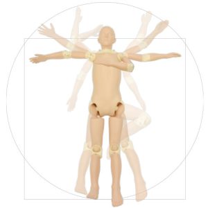
- Each joint has the close-to-human range of motion and can be positioned according to the target part of the shot.
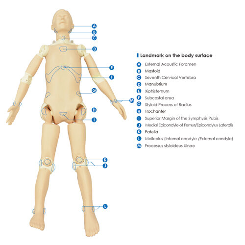
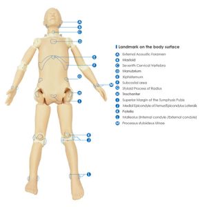
- Contains all necessary landmarks for positioning
- Specialized in positioning and has been drastically made lighter (18kg).
- Enables training free from privacy concerns and inconveniences associated with use of standardized patients.
- No metal parts, which causes artifacts, are included in the phantom.
Training skills / Applications
- Patient positioning
- Patient transportation
- Plain radiography
Case / Pathology
- [Skeleton] Skull, cervical spine, vertebrae, clavicles, scapulae, sternum, pelvis, lungs (without vessels), heart, kidneys, upper and lower arm bones, carpal, metacarpal, femur, kneecaps, lower leg bones, tarsi, metatarsals, phalanges.
- [Internal organs] trachea (up to 1st bifurcation), lungs (diaphragm only), heart, kidneys
Size (approx)
- Chest girth: 85cm (body thickness: 20cm)
- Waist girth: 75cm (body thickness: 19cm)
- Height: 165cm
- Weight: 18kg
Application
- Bone scintigraphy
- Bone SPECT
- CT
- NaF-PET
Materials
Soft fabrics:
- polyurethane foam (density 0.2)
Skeleton:
- epoxy resin (density 1.31
Skull:
- urethane resin (density 1.12)
| Quantity | Item |
|---|---|
| 1 | Adult Whole Body Phantom |
| 1 | Head Stand |
| 1 | Tools for Assembly |
| 1 | Radiography Data |
| 1 | Clothes |
| 1 | Instruction Manuals |
*Separable into 10 parts
Radiographic Condition
Because it is designed with a focus on positioning, the imaging conditions are not the same as clinical conditions for imaging of the human body. In order to reduce the operator’s radiation exposure and the stress to the device, the phantom is designed for imaging using only from one-half to one-third of the average radiation used under normal clinical conditions.
Landmarks for Positioning
External Acoustic Foramen / Mastoid / Seventh Cervical Vertebra / Manubrium / Xiphisternum / Styloid Process of Radius / Superior Margin of the Symphysis Pubis / Medial Epicondyle of Femur/Epicondylus Lateralis / Patella / Malleolus (Internal condyle /External condyle) / Subcostal area / Landmark on the body surface / Trochanter / Processus styloideus ulnae

Realizing Education That Inspires Through Manufacturing
Founded in 1891, Kyoto is the world’s only manufacturer of anatomical models, simulators, and imaging phantoms. Made in Kyoto, Japan and trusted in over 60 countries around the world. Passing on cultural heritage through technologies for restoring and replicating cultural assets.