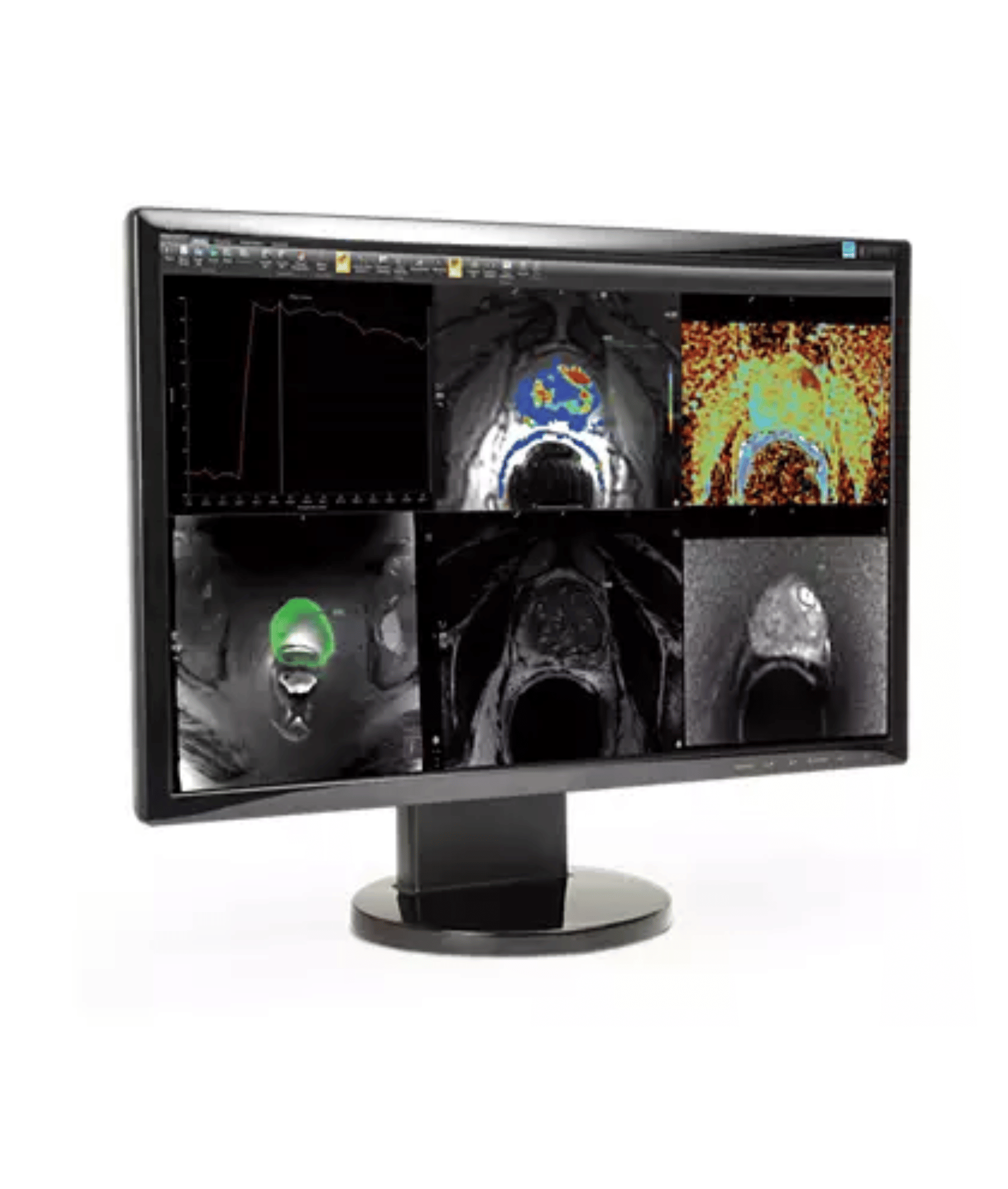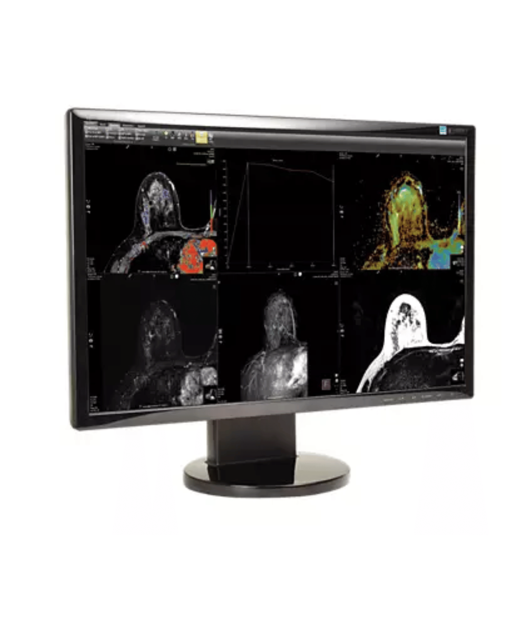

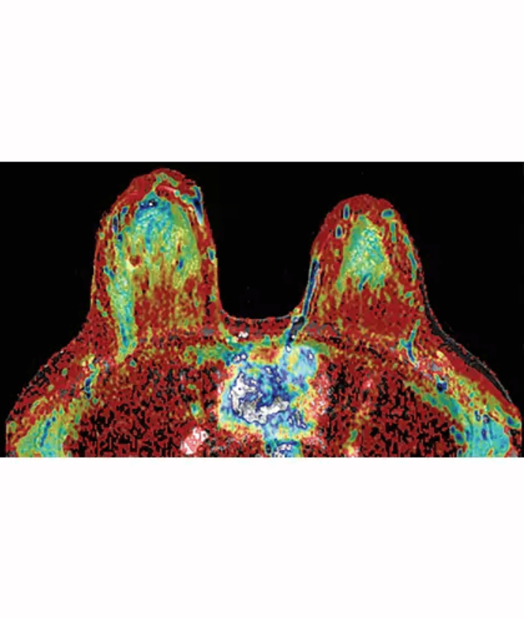
Customizable displays of post-processed data
Take advantage of an advanced post-processing engine that automatically generates multi-planar reformatted (MPR) and maximum intensity projection (MIP) images. You can also apply 3D image registration to correct motion artifacts, and chest wall/cardiac masking to help focus on the target anatomy. Additional tools are available to help analyze the data, such as subtraction images and color overlays based on kinetic characteristics. You can view the post-processed data in a customized hanging protocol or automatically forward it to a PACS archive.
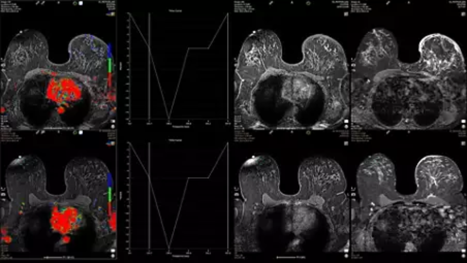
Convenient current/prior review facilitates comparative analysis
DynaCAD Breast automatically associates prior exams with current ones for quick and effortless side-by-side review. You can set linked exams to scroll sequentially, so you can visually compare current and prior images on a slice-by-slice basis.
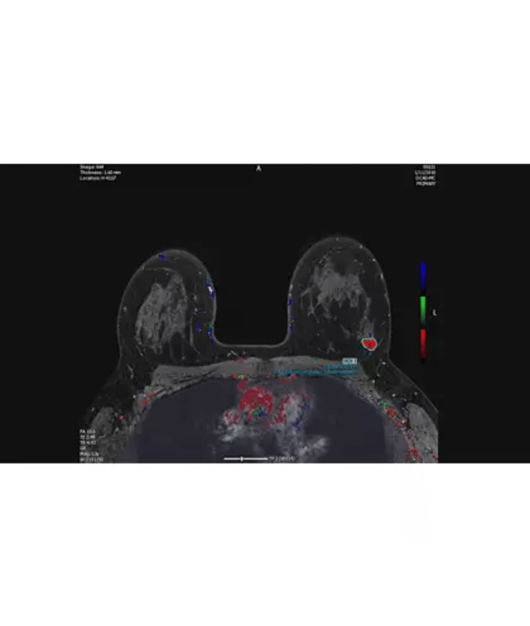
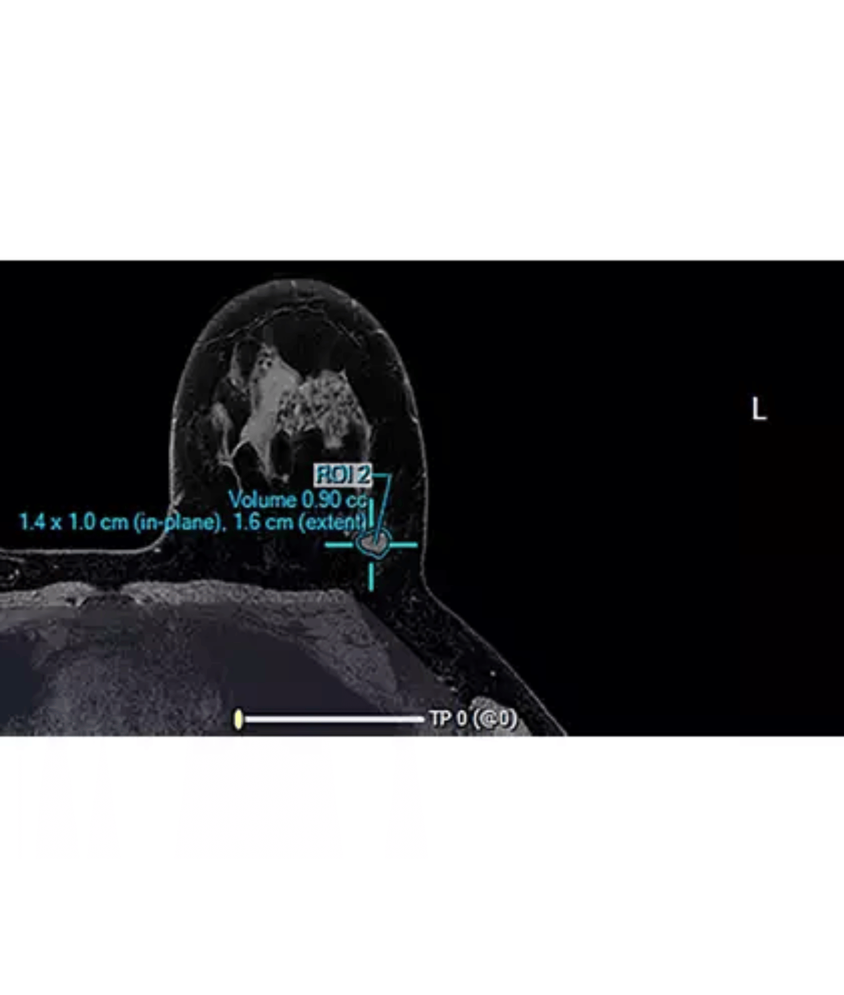
Lesion segmentation enhances workflow efficiency
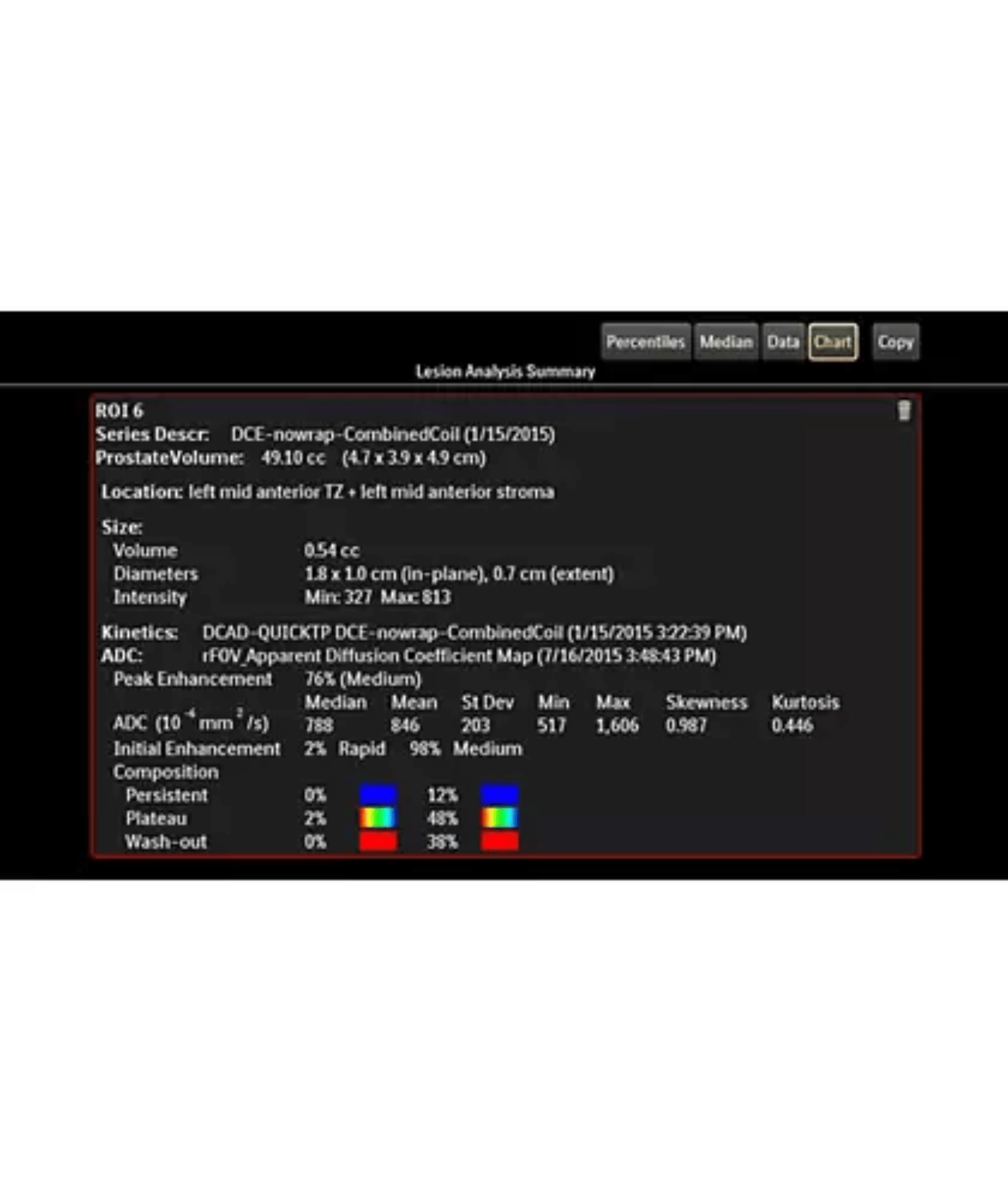
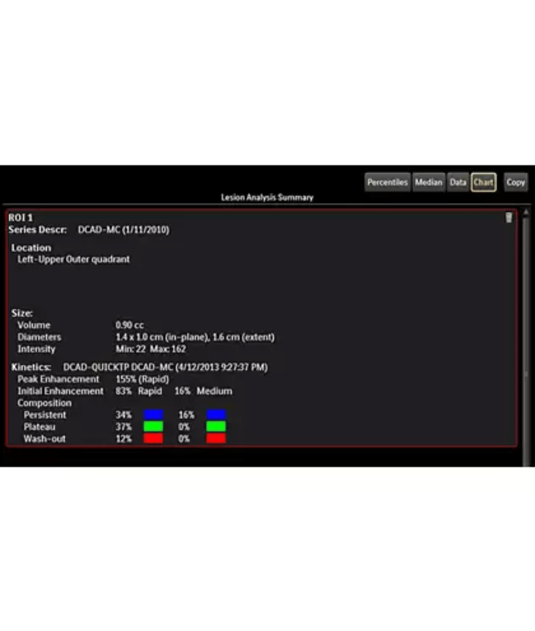
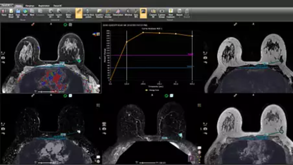
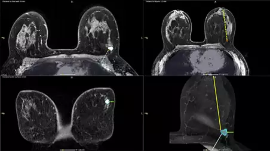
Launch an automatic segmentation feature with one mouse click. The advanced segmentation algorithm allows for your on-the-fly modification and presents you with a volume analysis, lesion composition statistics, histograms, and a 3D-rendered morphological overview. Auto-populated regions of interest (ROIs) can be viewed as a 2D image or can be mapped onto a maximum intensity projection, where it will appear as a 3D object. The resulting segmentation report provides a calculation of lesion location diameter measurements and location.
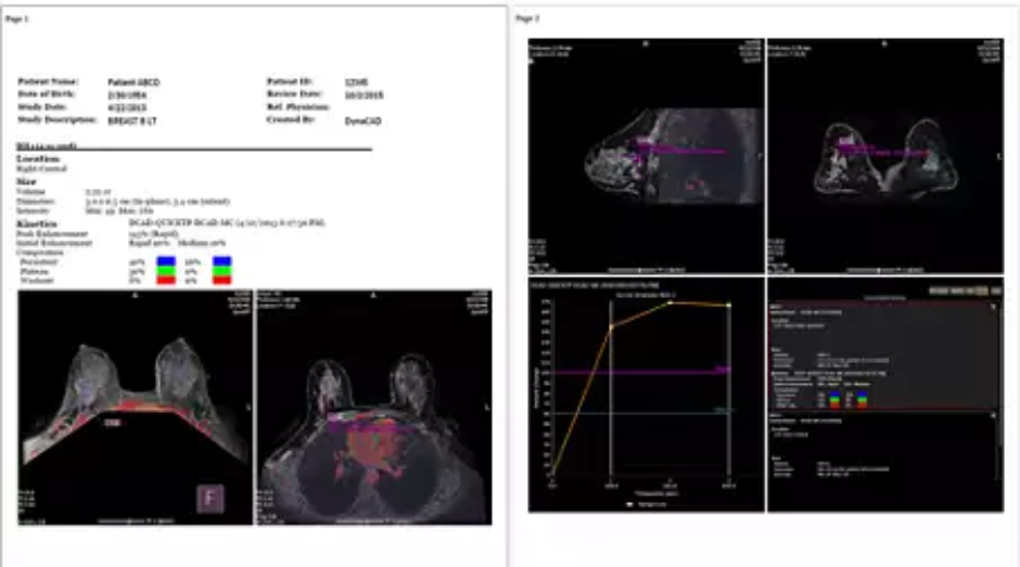
Automated reports capture and share relevant data
Use the structured reporting system in DynaCAD Breast to produce highly customized exam reports. You can program reports to automatically append pre-selected images containing kinetic data, measurements, and annotations. The system also automatically populates additional report fields such as lesion diameter measurements, lesion to landmark distances, and volumetric data. Upon completion, you can print patient reports, save as a PDF, or send as DICOM images.
Clearer images for more productive study reviews
Motion Correction helps reduce unwanted image artifacts, boosting time efficiency and confidence as studies are interpreted. The system can display corrected images by default, or users can toggle them on and off.
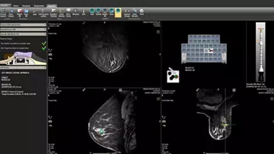
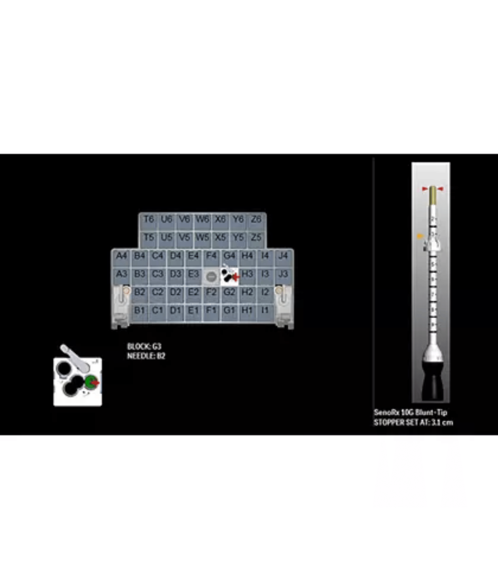
Enhanced workflow for biopsy procedures
DynaCAD Breast includes DynaLOC visual guidance for planning of breast biopsy procedures. DynaLOC’s menu-driven interface guides you through the initial setup of equipment to the confirmation of targets. Access a large library of interventional instruments and hardware for a custom, site-specific workflow. Computer renderings illustrate patient position, device setup, target area and needle tract for added confidence.