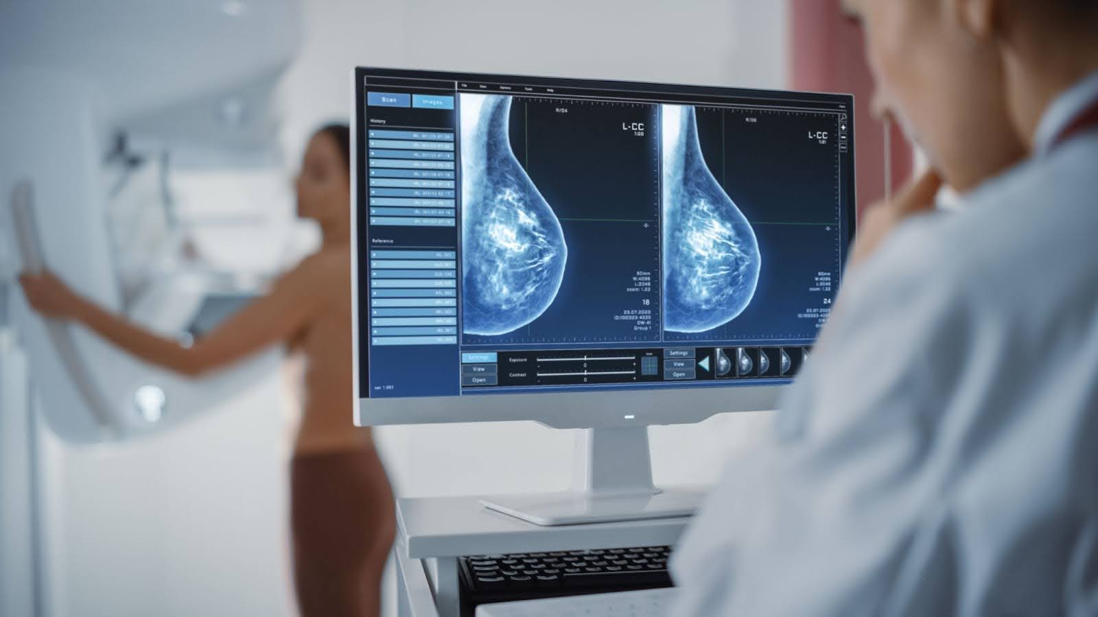
In mammography, clarity is not only about image quality. It is also about context.
Technologists regularly encounter skin features, scars, or areas of patient concern that may appear significant on imaging but are clinically benign. Without clear identification at the time of acquisition, these features can resemble pathology, introducing uncertainty during interpretation and sometimes leading to additional imaging or unnecessary patient concern.
Purpose-built mammography markers provide a simple way to document these details directly within the image, ensuring that what the technologist observes during positioning is clearly communicated to the radiologist during reporting.

When Visual Context Matters
Breast imaging relies on subtle differences in tissue appearance. Small anatomical variations can look markedly different once compressed and imaged, and written notes alone rarely provide the same clarity as a visual reference captured within the study itself.
Markers allow areas such as moles, scars, nipples, or palpable concerns to be clearly identified at the time of imaging. By distinguishing normal external features from internal findings, they help reduce ambiguity and support more confident interpretation. This becomes particularly important in screening environments, where efficiency and diagnostic certainty must exist side by side.
As imaging technology becomes more sensitive, the ability to clearly communicate clinical context within each image becomes increasingly valuable.

Designed for Modern Mammography and Tomosynthesis
Mammography SPOT® and TomoSPOT® markers are designed specifically for digital mammography and breast tomosynthesis systems. Rather than obscuring anatomy, the markers are engineered to remain visible while allowing underlying tissue detail to be assessed clearly.
These markers assist clinicians by clearly identifying anatomical landmarks and areas of concern while maintaining diagnostic visibility. Their low-density, non-metallic construction ensures compatibility with modern imaging systems without introducing distracting artefacts or compromising image quality.
Different marker shapes provide immediate visual meaning within the image, allowing radiologists to quickly understand whether a feature relates to a mole, scar, nipple, or palpable finding without needing additional clarification.
Supporting Confidence Across the Imaging Team
Although small in size, mammography markers influence multiple stages of the imaging process. For
technologists, they provide reassurance that areas discussed with the patient are accurately documented. For radiologists, they offer reliable reference points that support efficient and confident interpretation.
Patients benefit as well. Clear identification of benign features can reduce the likelihood of additional views or recall appointments, helping minimise anxiety during what is often a stressful screening experience.
Over time, consistent use of markers becomes less of an added step and more a routine part of producing complete, communicative imaging studies.
A Practical Addition to Breast Imaging Workflows
As breast imaging continues to advance, precision extends beyond resolution and detector technology. Small workflow tools that improve communication between acquisition and interpretation play an equally important role.

Mammography markers provide a straightforward way to preserve clinical context within each study, supporting clearer reporting while maintaining efficient workflow in busy imaging environments.
If you’re reviewing breast imaging consumables or standardising markers across your department, visit our website or view the Imaging Markers and Procedure Accessories catalogue to explore the available range.