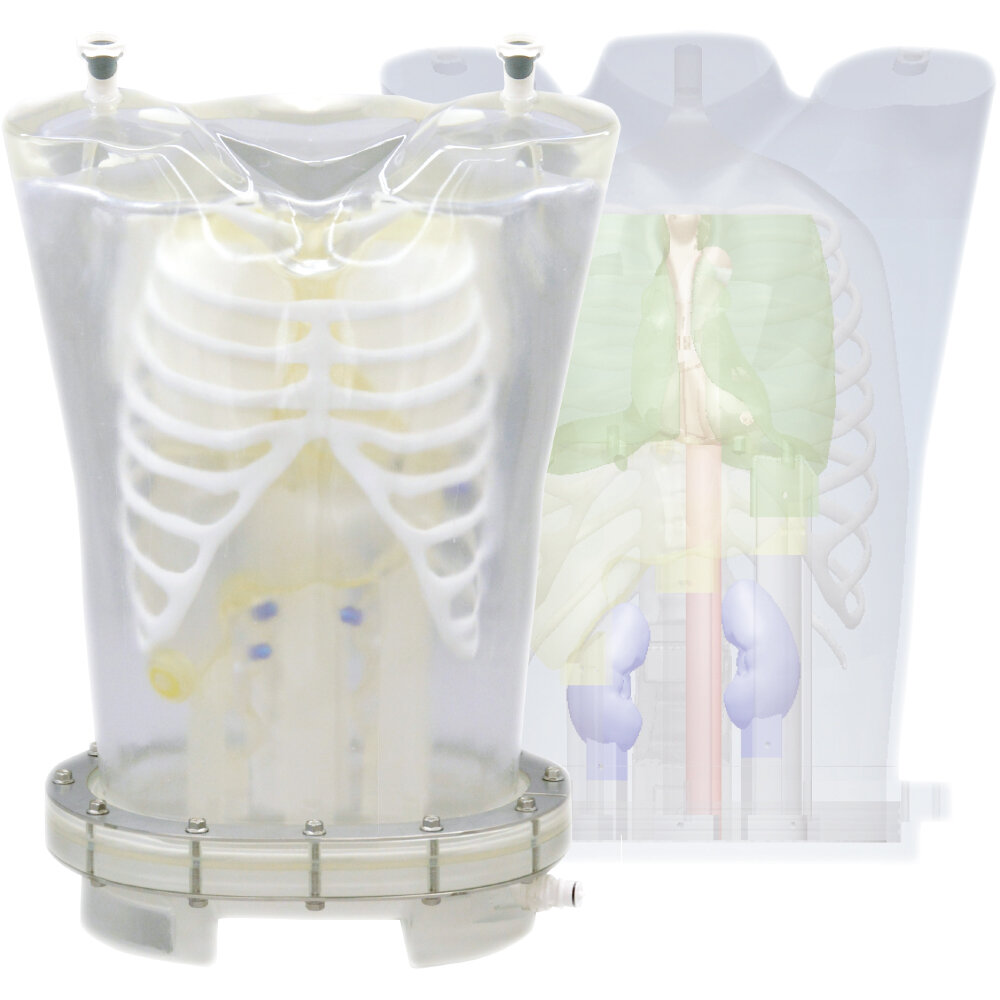
Features
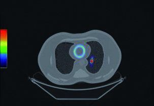
Examination of myocardial density through SPECT imaging
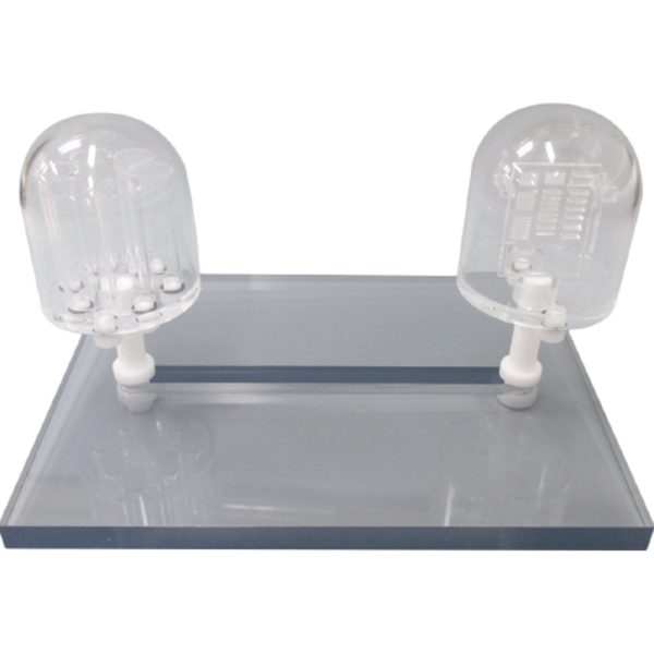
- Verification of myocardial imaging with the use of various RI solution densities
- Ability to capture defects of the myocardial region
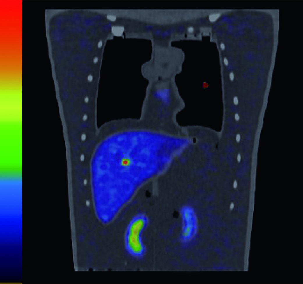
- Can reproduce image variations of the heart by injecting RI solutions in the liver, kidney and lungs
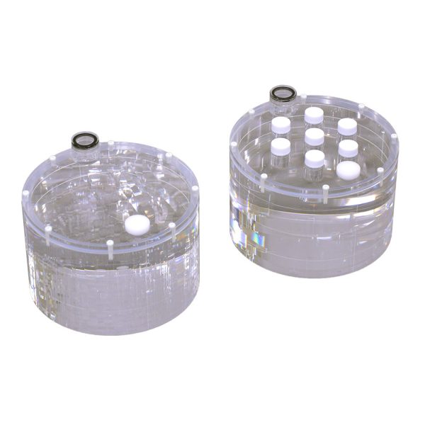
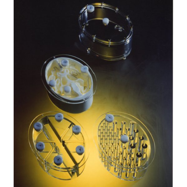
Examination of RI solution density for simulated tumors
- The simulated tumors can be inserted into lung, liver and breast
- Tumors can be filled with FDG/RI solution into the spheres for evaluation of density, size and placement
Applications
- PET/SPECT
- Quality management of NM equipment
- Myocardial density with SPECT imaging
- RI solution density for tumor imaging
Materials
Soft tissue: transparent polyurethane
Lungs: materials with density 0.4 g/cm3
Bone materials: Calcium-infused material to provide proper attenuation with use of RI solutions
Anatomy
- Liver
- Lung (right/left)
- Kidney (right/left)
- Hot spots (liver, lungs and breast)
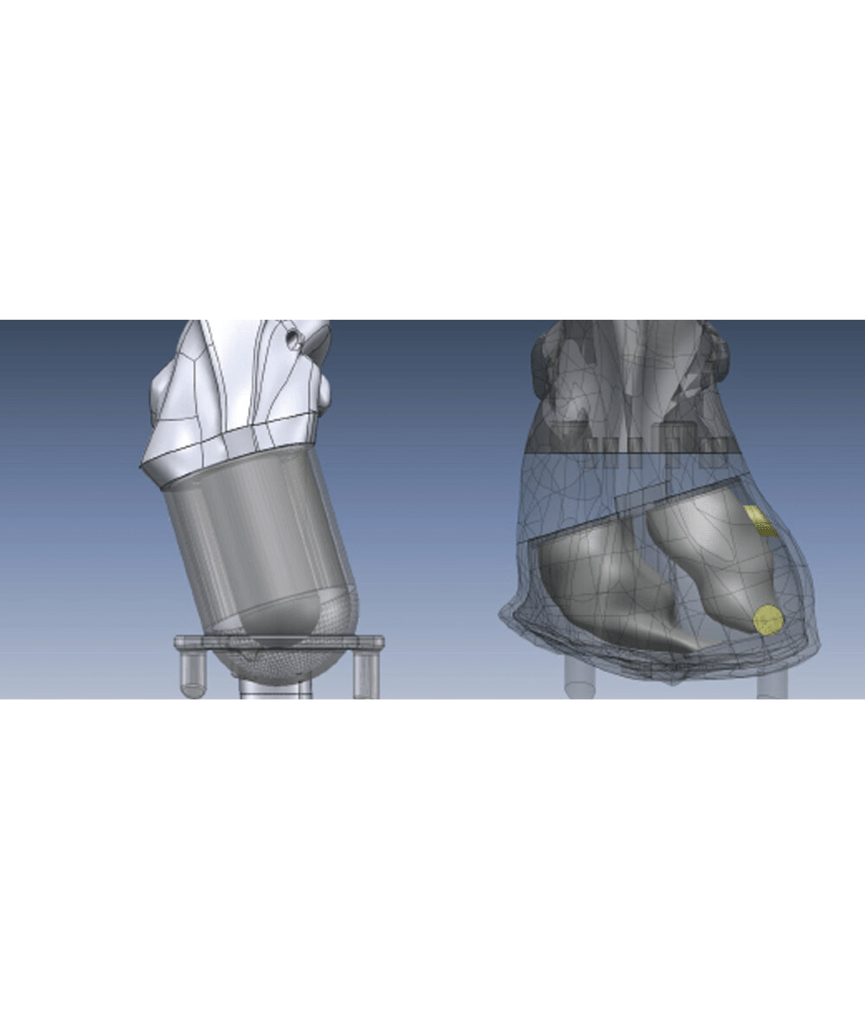
- Heart
Anatomical type: right ventricle, left ventricle and myocardium
Geometric type: left ventricle and myocardium
- Bone: 370HU
- Lung: -900HU
- Organ shell material: 100HU, and 1.16g/cm3 in density
Phantom size
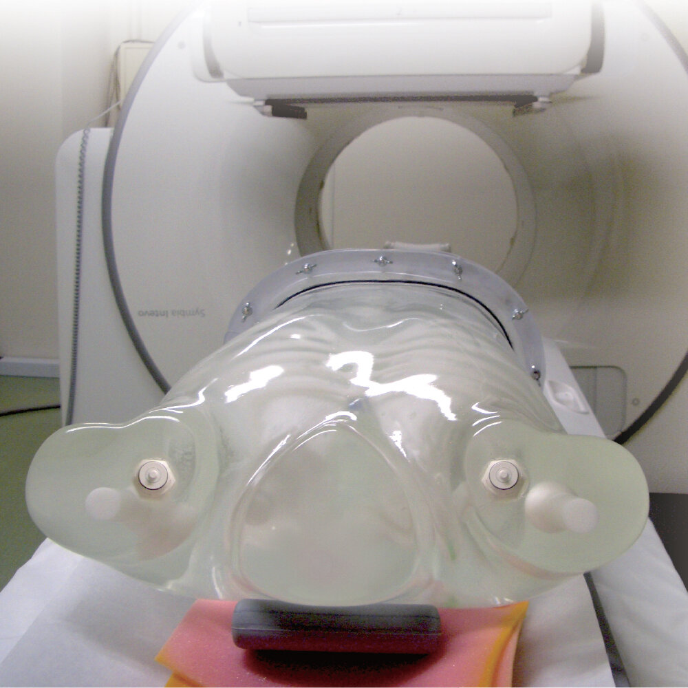
W44×H69.4 cm
(W17.3×H27.3 in)
Phantom weight
- phantom itself: 21 kg / 46.2 lb
- when filled with liquid: 37.5 kg / 82.6 lb
Set Includes
| Quantity | Item |
|---|---|
| 1 | Thorax Body |
| 1 | Base |
| 2 | Lungs (Left and Right) |
| 6 | Plastic Pins |
| 4 | Hearts |
| 6 | Supporting Bars |
| 1 | Liver |
| 4 | Metal Frames |
| 2 | Kidneys (Left and Right) |
| 1 | Silicon Sealing Gasket |
| 1 | Rib Cage and Spine |
| 3 | Tubes |
| 2 | Breasts (Left and Right) |
| 1 | Syringe |
| 3 | Hot Spots |
| 12 | Nuts and Bolts |
| 2 | Aorta |
| 1 | Water Tank |
| 1 | Beaker |
| 1 | Vaseline |
| 1 | Manual |

Realizing Education That Inspires Through Manufacturing
Founded in 1891, Kyoto is the world’s only manufacturer of anatomical models, simulators, and imaging phantoms. Made in Kyoto, Japan and trusted in over 60 countries around the world. Passing on cultural heritage through technologies for restoring and replicating cultural assets.