")
Description
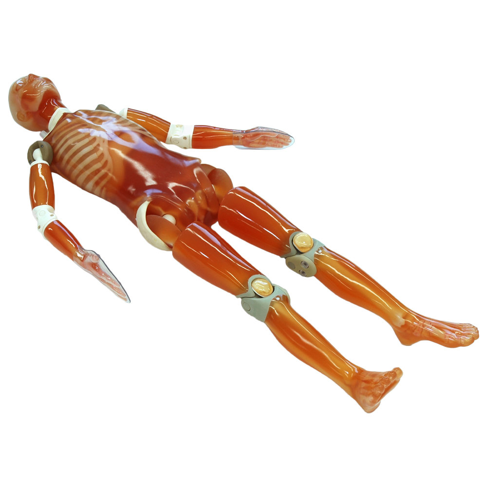
TAKE-APART PIXY WITH LUNGS, HEART, LIVER, URETERS, LARGE INTESTINE, CIRCLE OF WILLIS, AND ESOPHAGUS
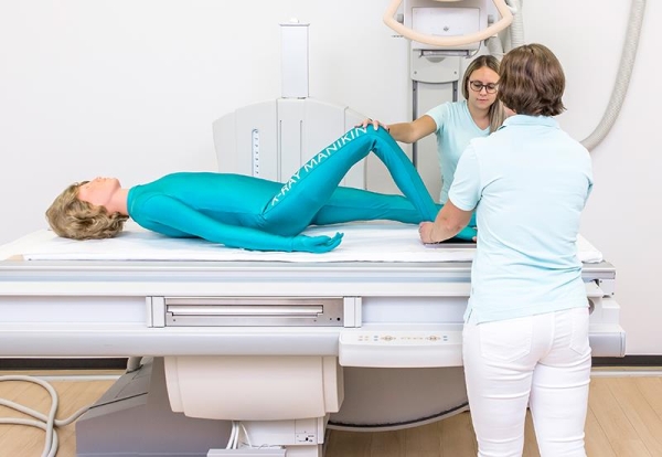
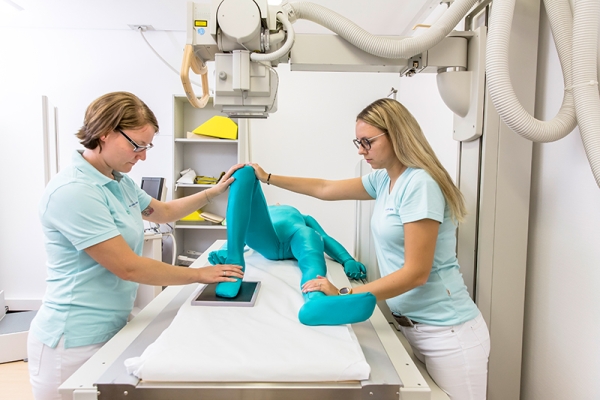
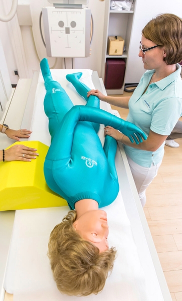
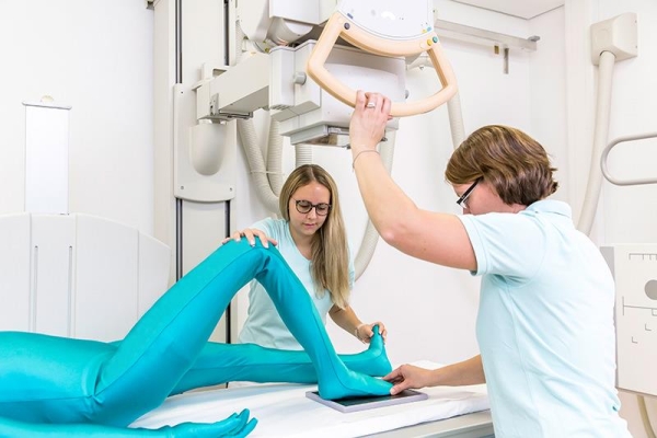
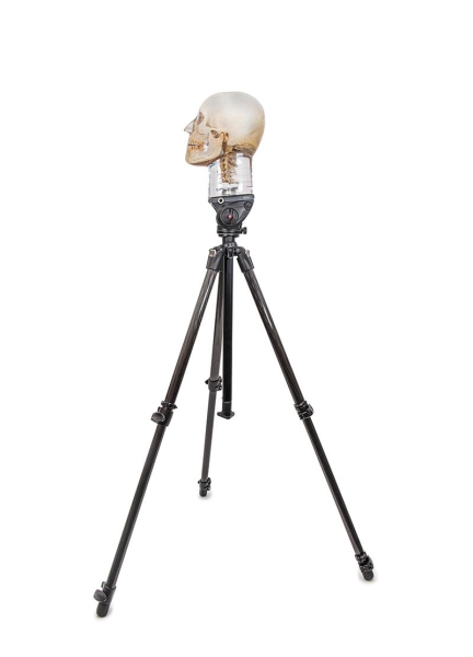
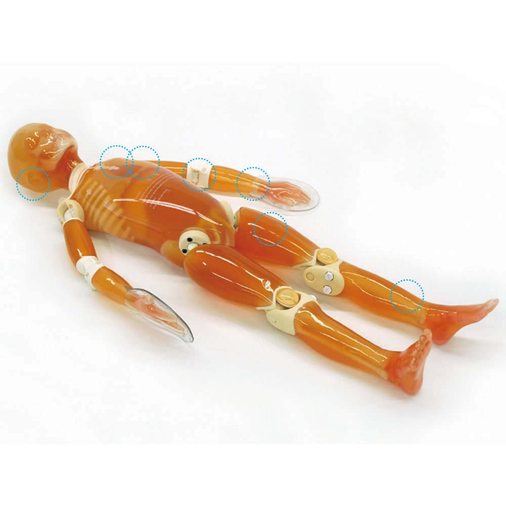
Take-Apart CT Pixy is an anatomically and radiologically correct female designed specifically for training radiologic technologists. At 5’ 1” (156 cm) weighing 105 lbs (48 kg), Take-Apart CT Pixy is a repeatable, convenient substitute for patients and virtually indestructible.
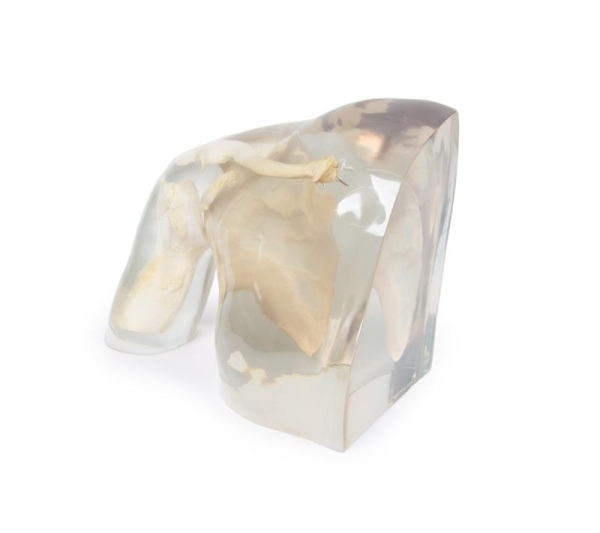
Take-Apart CT Pixy may be ordered with or without fill ports. In addition to the stomach, gall bladder, urinary bladder, kidneys, rectum, and sigmoid flexure, Take-Apart CT Pixy additionally includes lungs, heart, liver, ureters, large intestine, Circle of Willis, and esophagus. Custom pathologies and traumas are available at an additional cost.
Built with soft-tissue mold and skeleton molds that are matched for anatomic fidelity, Take-Apart CT Pixy permits unlimited exposures, demonstrates the effects of changing technical factors, and allows for the evaluation of student performance. Students have no difficulty in maneuvering Take-Part CT Pixy into the most desired positions as the phantom is built to tolerate trainee errors.
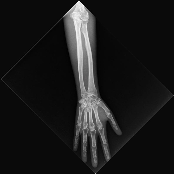
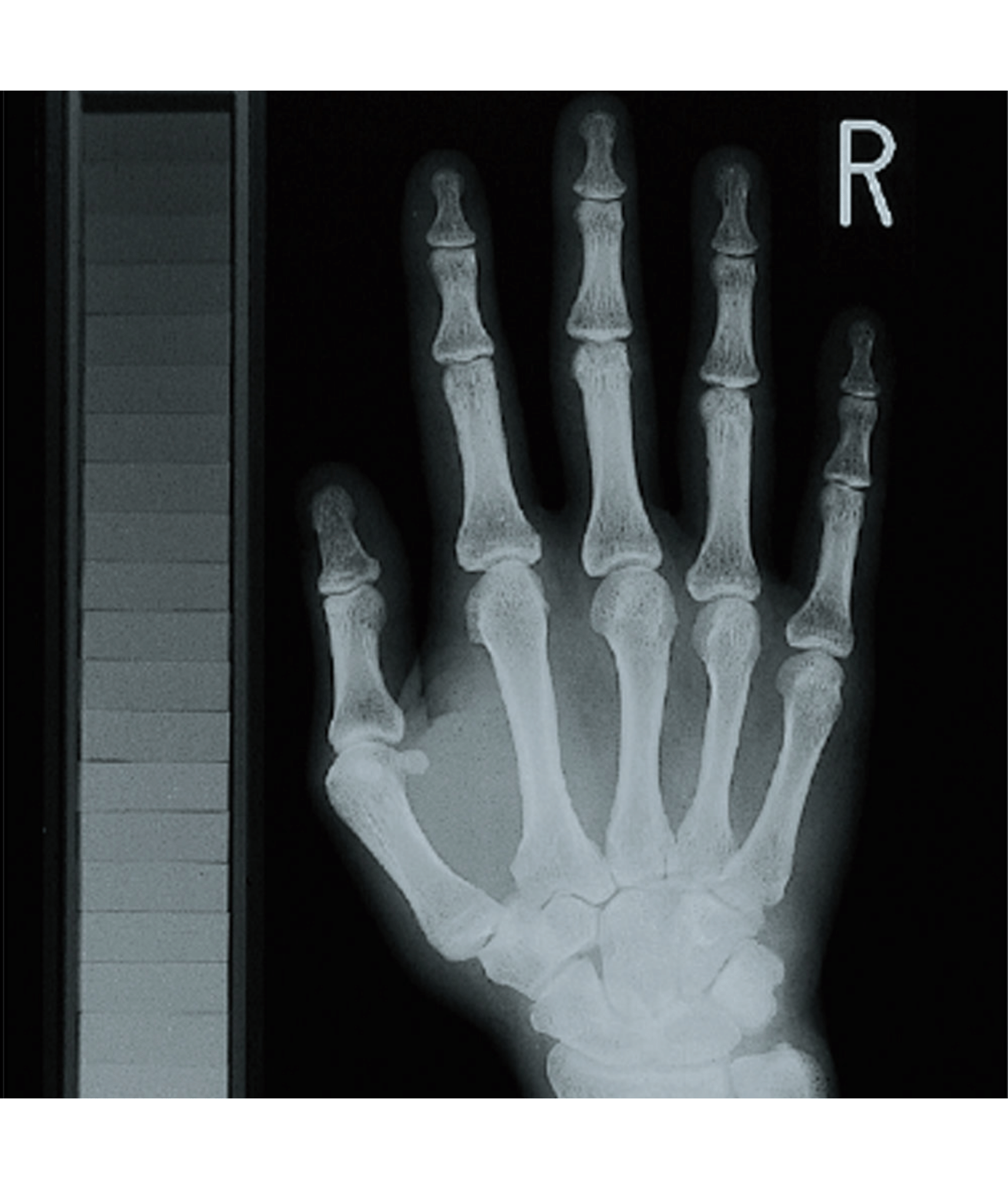
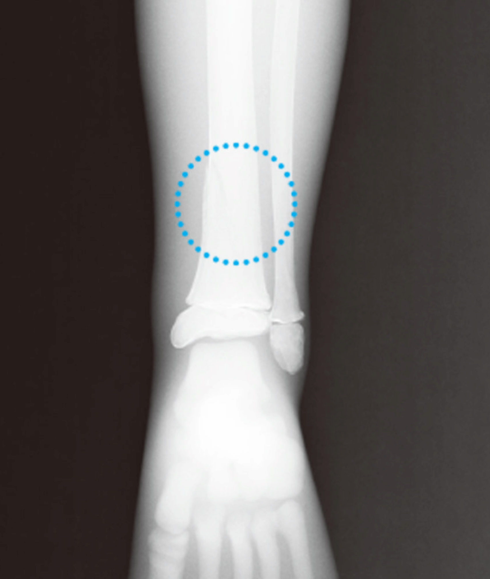
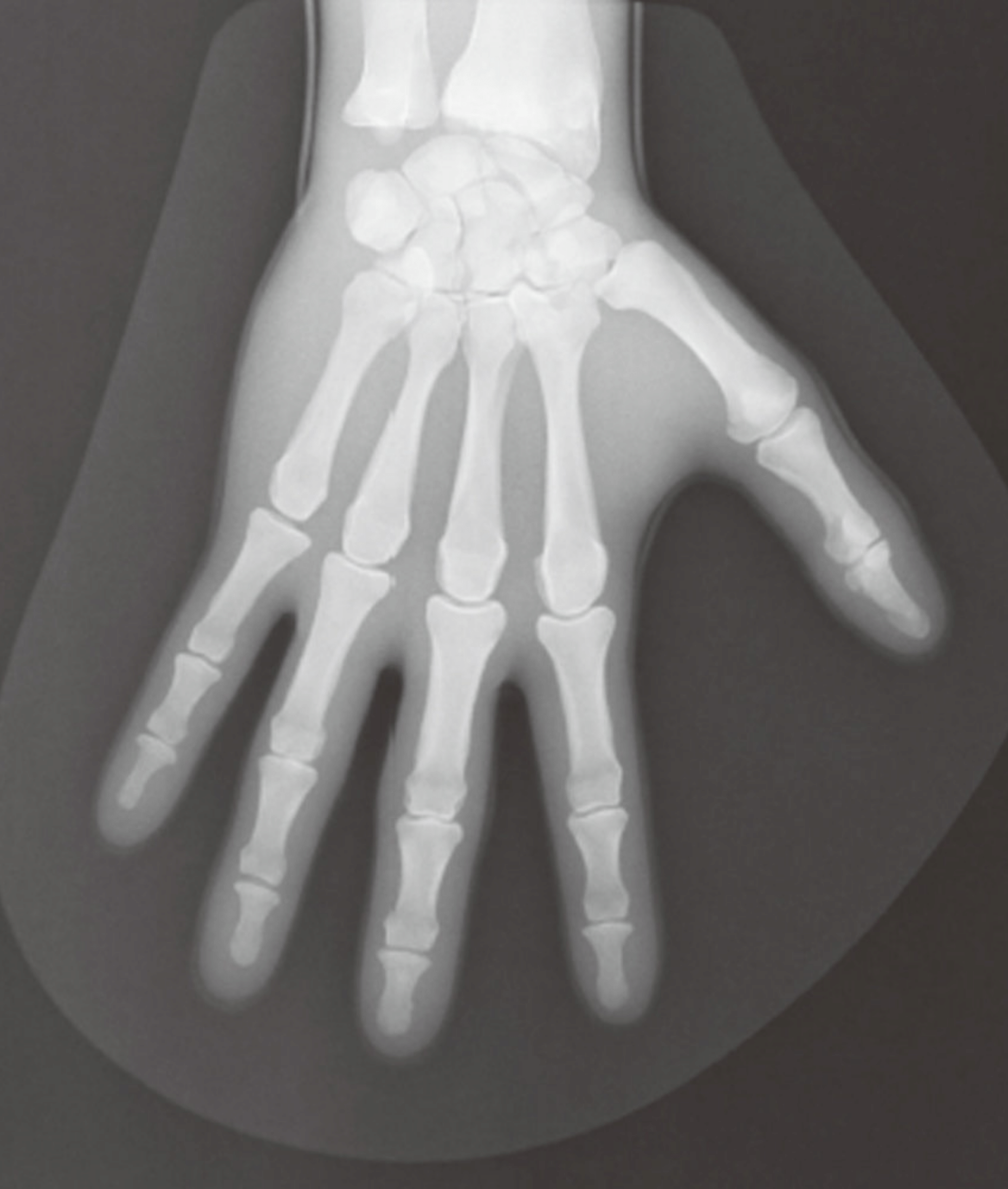
Take-Apart CT Pixy is used to demonstrate anatomy and evaluate positioning and imaging techniques, including kVp, mAs, contrast, optical density, digital processing, OFD, and TFD. Made of tissue-equivalent materials and life-like articulations, Take-Apart CT Pixy is more realistic than a cadaveric skeleton with radiographs that are optically equivalent in density and contrast to human patients.
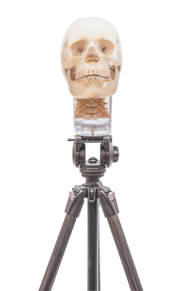
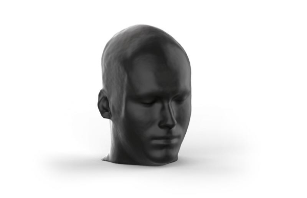
C1, C2, C6, and C7 were converted to mechanical nylon joints because educators in the field prefer full positioning capabilities for the head. This design permits the remaining neck vertebrae to be fixed in a normal position while assuring a full range of head motion.
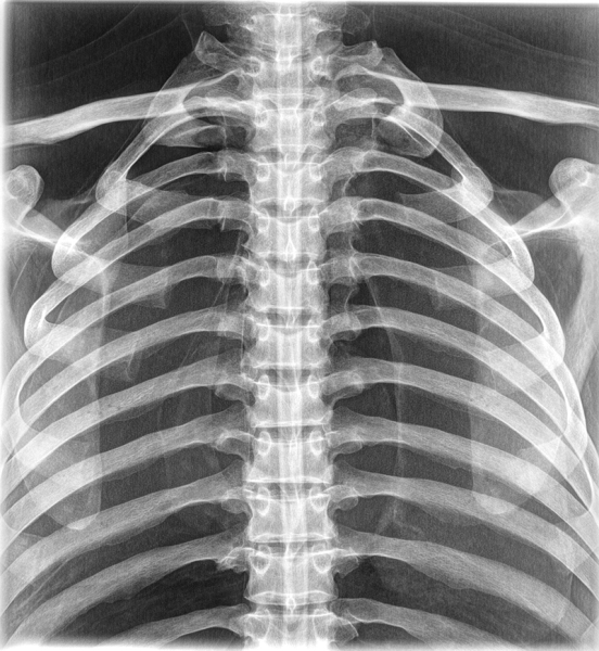
The skull of Take-Apart CT Pixy has frontal and sphenoidal sinuses, ethmoidal and mastoid air cells, and the auditory ossicles. Bone sutures are radiographically visible.
Take-Apart CT Pixy lungs are molded of tissue-equivalent foam with the mass density of inflated human lungs (0.30 g/cc). They are connected to the oro-nasal cavity by the stem bronchi and trachea. The oro-nasal pharynx is filled with a nearly air-equivalent foam.